
Beyond the Brain: Unveiling the Mind-Muscle Connection with Simultaneous NIRS
Near-infrared spectroscopy (NIRS) can be applied on any tissue to measure local oxygenation. In an increasing number of studies, NIRS is applied on the brain and muscle simultaneously to get information on neural and muscular activity during for instance exercise, exposure or intervention. In this blog post, we describe application areas with frequent use of simultaneous NIRS brain and muscle measurements, highlight studies per category and explain how our products can provide the optimal solution for combined measurement.
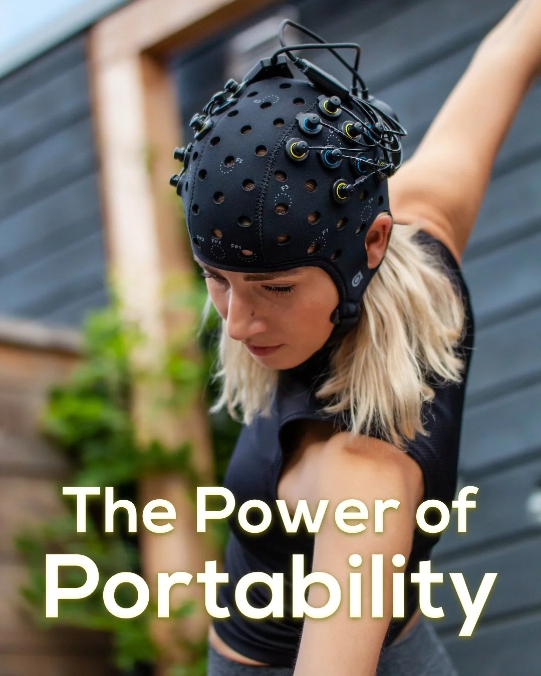
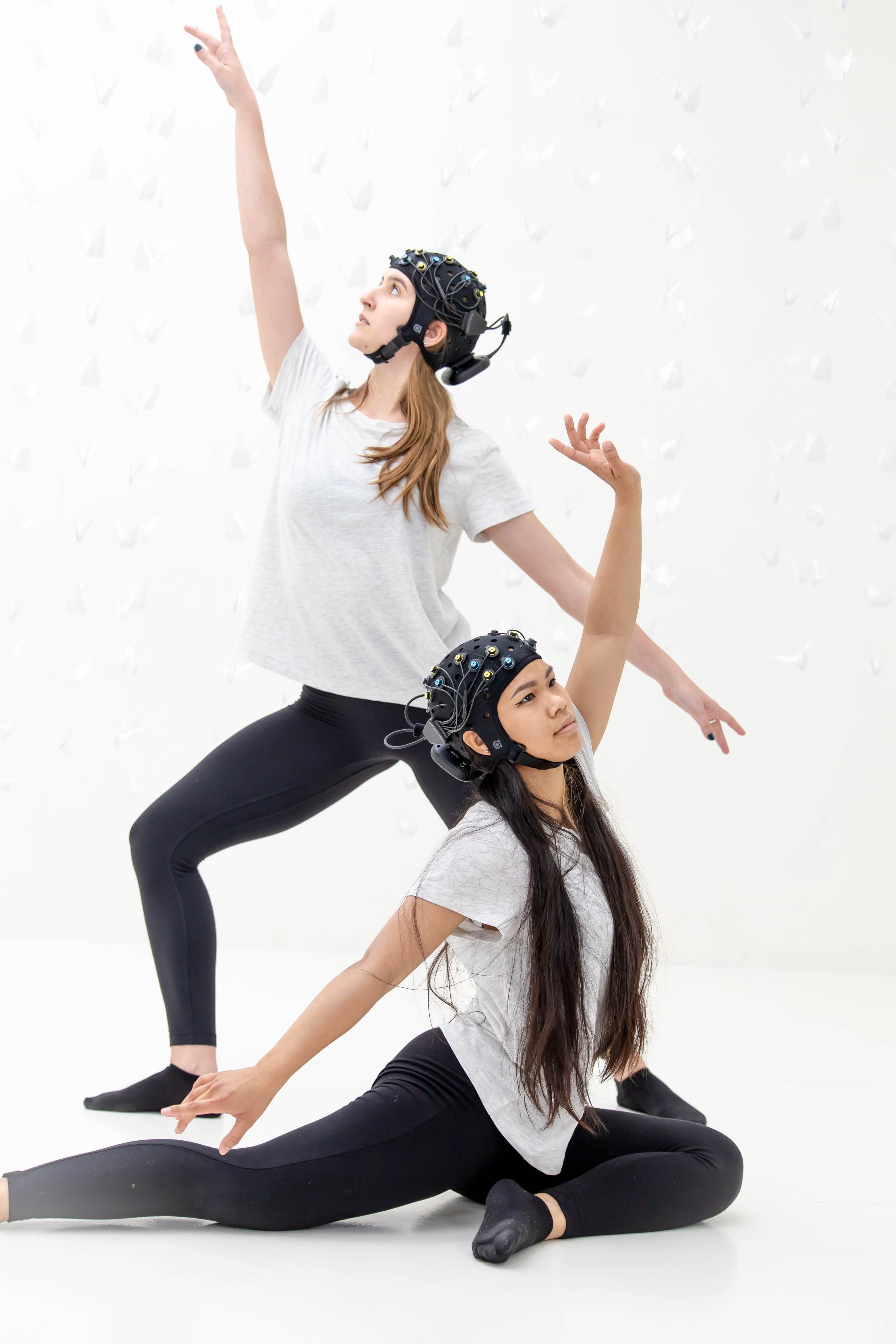
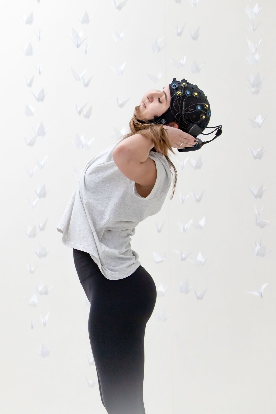
Near-Infrared Spectroscopy (NIRS) is a non-invasive technology that measures relative concentration changes in oxygenated hemoglobin (O2Hb) and deoxygenated hemoglobin (HHb) in any local tissue, for instance muscle or brain. As it is non-invasive, easy to use and often portable, NIRS can be applied in a huge range of research fields, even including experiments that involve movement or exercise. Its advantages make it an ideal tool to be used not only for experiments performed in the lab, but also in more naturalistic settings.
Application possibilities for simultaneous NIRS brain and muscle measurements
In an increasing number of application areas, NIRS is measured in brain and muscle simultaneously is to achieve a more complete picture of systemic oxygenation in the body. In the following, we explain potential benefits of combining brain and muscle NIRS per category, and highlight important recent publications.
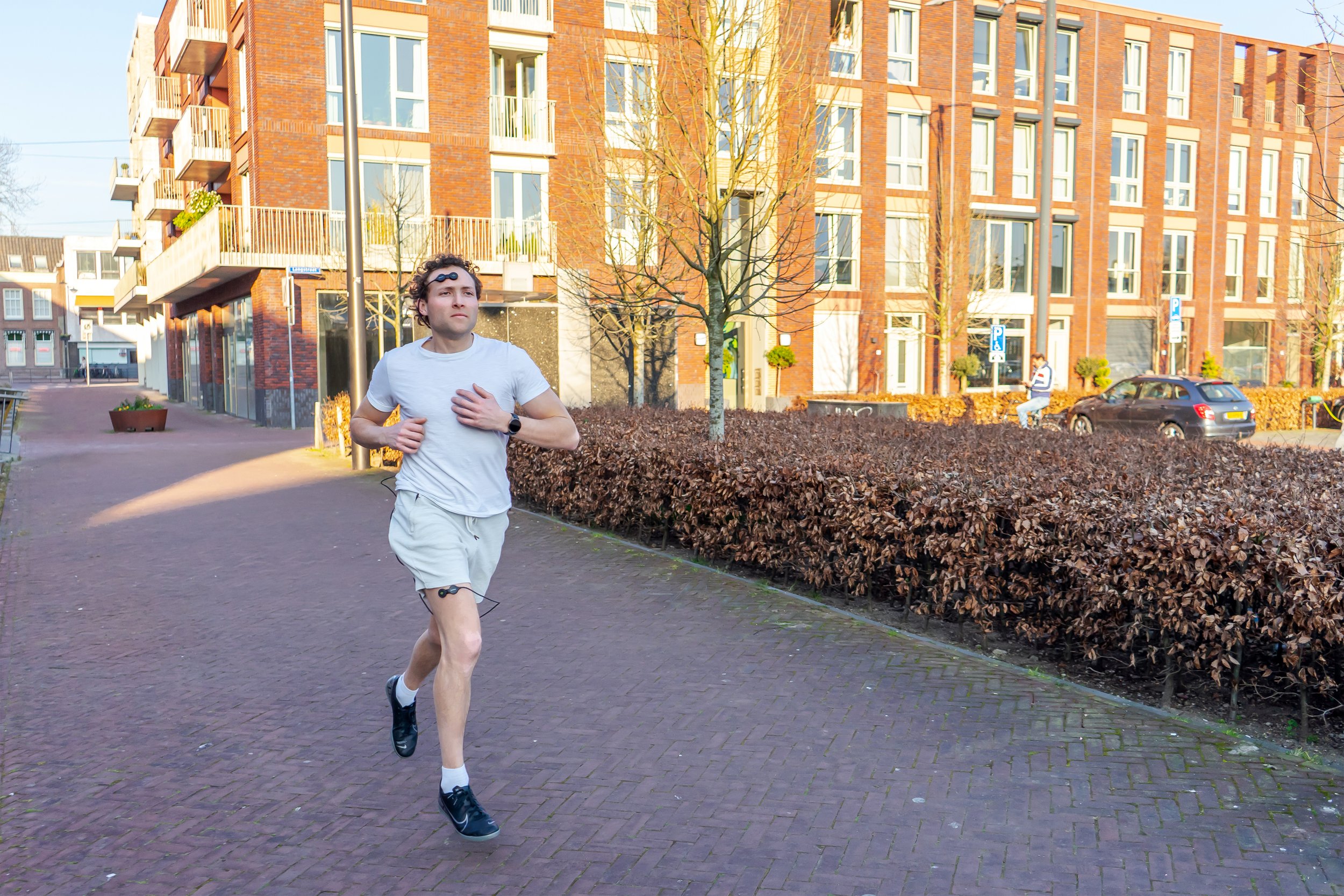
Sports Science - Comparing muscle and brain oxygenation
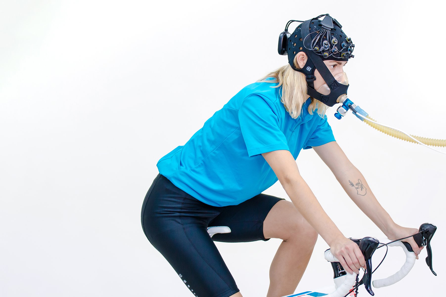
NIRS found its first applications often in sports science studies, especially to monitor local muscle oxygen consumption in various muscles. In recent years, using fNIRS to measure brain activity is increasingly used in Sports and Exercise Science, as it can give information on cognitive state, decision-making processes, or motor activity during exercise. Hence, fNIRS is predominantly applied in the prefrontal and/or motor cortex. Measuring muscle and brain oxygenation simultaneously during exercise can be used for different reasons: It can help to achieve a complete picture of systemic oxygenation, compare neural and muscular oxygen status, or assess the effect of certain training strategies to improve sports performance.
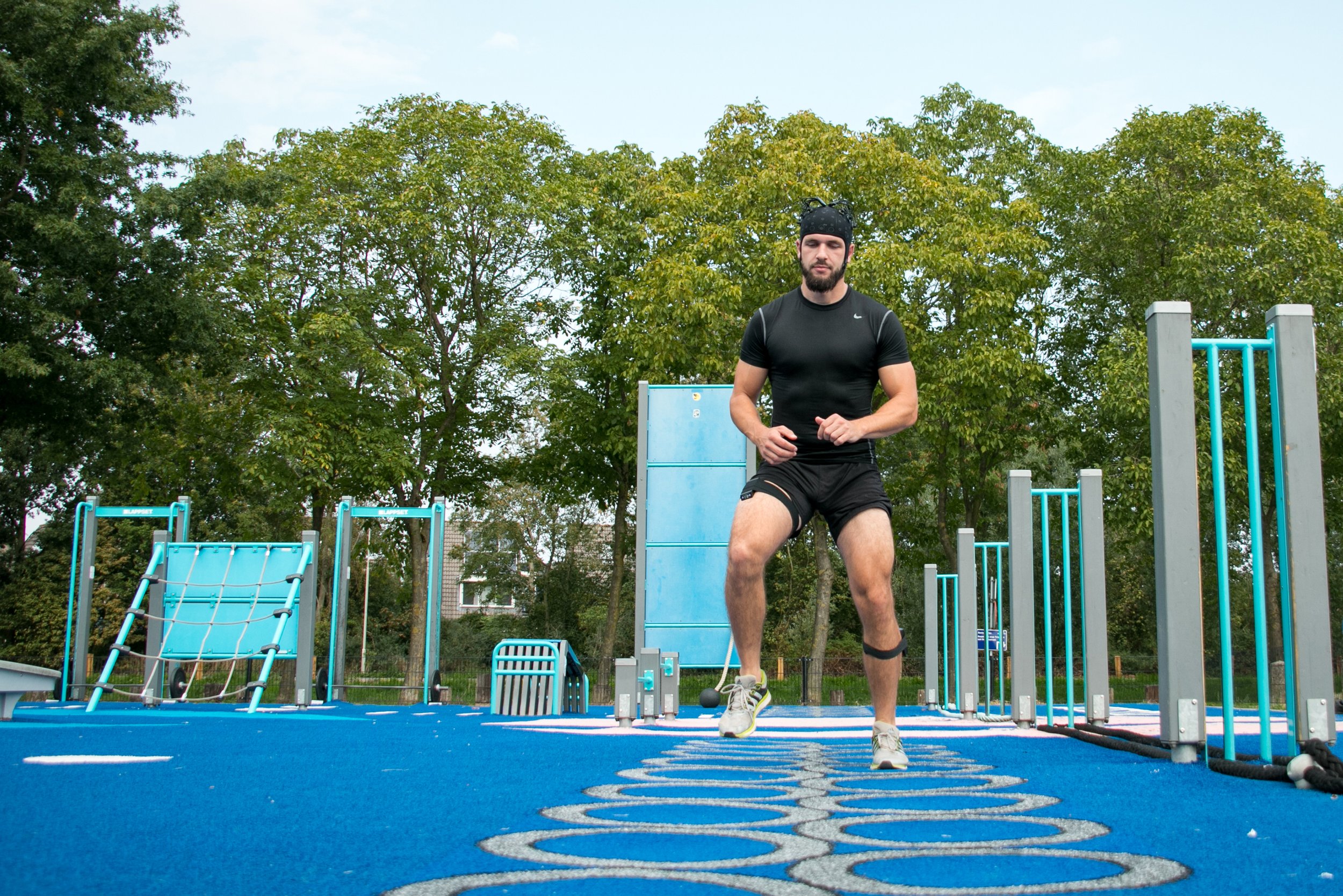
Zadeh et al. performed a study to simultaneously measure and compare neural and muscular oxygenation during progressive exercise. Participants performed maximal incremental exercise test, while NIRS was measured on the prefrontal cortex and vastus lateralis with the PortaLite / PortaMon, before and after the anaerobic threshold (AT) was reached. In the vastus lateralis, Hb-diff (difference between O2Hb and HHb) significantly decreased after AT. On the contrary, an increase in Hb-diff in the prefrontal cortex after AT was reported, indicating well-functioning cerebral autoregulation even during metabolic fatigue. Findings verified the feasibility of measuring brain and muscle oxygenation at the same time with NIRS during progressive exercise.
In a study by Cherouveim et al., the association between muscle blood flow restriction, alterations in cerebral hemodynamics, and decline in exercise performance was assessed. Amongst others, the OxyMon was used to simultaneously measure prefrontal brain activity and muscle oxygenation on the vastus lateralis, while participants performed maximal incremental exercise with (WC) and without (NC) blood flow restriction. Muscle oxygenation and exercise performance significantly decreased in NC condition compared to WC. Significant lower brain oxygenation at maximum effort could be found in WC compared to NC condition.
Hypoxia - Achieving a complete picture of oxygenation in hypoxic conditions
Hypoxia is defined as any condition where the demand for oxygen by a tissue exceeds the actual oxygen supply. Understanding the physiological mechanisms in hypoxic conditions and which potential coping mechanisms the human body can develop to counter hypoxia is crucial. As NIRS can measure oxygenation status in local tissue, it has been successfully used as a tool in hypoxia and altitude research for many years.
Exposure to hypoxia or altitude can influence oxygenation in the complete body, but probably to different extents and time-delayed. Therefore, in recent years an increasing number of NIRS papers measuring the brain and muscle at the same time during hypoxia can be identified. Especially during exercise, hypoxia can alter oxygen supply and hence various studies assess exercise performance in hypoxic conditions.
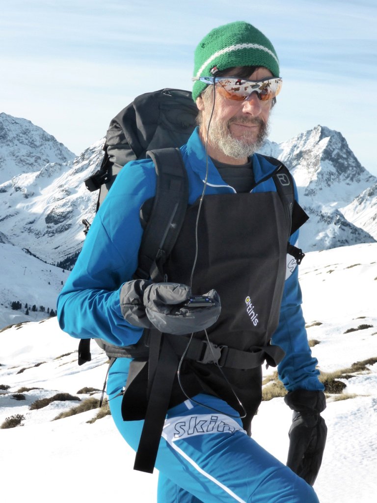
As an example, Rupp et al. compared the effects of normobaric hypoxia (NH) and hypobaric hypoxia (HH) on cerebrovascular and muscular responses during self-paced aerobic exercise using the OxyMon. HH resulted in increased impaired performance and pacing compared to NH, likely due to differences in systemic alterations and cerebral oxygenation. HH required higher adaptive cost for maintaining cerebral oxygen delivery, resulting in decreased absolute power output compared to NH.
Similarly, Rosales et al. assessed the effects of acute normobaric and hypobaric hypoxia on human physiology. The OxyMon was applied to both brain and muscle simultaneously, to achieve a complete picture of oxygen status during different conditions. In the muscle, HbO2 decreased and HHb increased during exercise in all conditions. Brain oxygenation was lower in high altitude hypoxia compared to sea level. The study indicates that during hypoxia, muscle can keep oxygen status, but brain does not, which might be caused by cardiac compensation dependent on the environment. Hence, caution when equating different types of hypoxic exposures is required.
Baur et al. performed a study to investigate, if load carriage in normoxia and hypoxia alternates hemodynamics, tissue oxygenation and metabolism, as well as ventilatory responses. Participants performed baseline and experimental exercise trials during three different conditions. Amongst others, OctaMon and PortaMon were used to simultaneously measure frontal brain hemodynamics, and skeletal muscle oxygenation on vastus lateralis, respectively. Results demonstrated reduction of cardiorespiratory efficiency and increase of strain with load carriage especially during hypoxia, which might lead to increased risk for altitude sickness.
Clinical and Rehabilitation - Enhancing effectiveness assessment of therapy and rehabilitation
NIRS is frequently used in clinical research to assess muscle or brain oxygenation in different application areas, such as cardiovascular or neurological studies. Simultaneous measurements of brain and muscle are relatively new in clinical and rehabilitation experiments but can offer certain advantages, for instance receiving additional information on the effectiveness of therapy or rehabilitation strategies in different diseases and patient groups.
To this end, Dutta et al. performed a study to assess the potential of neuromuscular electrical stimulation (NMES) as a possible therapy tool in patients suffering from chronic venous insufficiency (CVI). Muscle NIRS was measured on the calf muscle, and simultaneously, the OctaMon was applied to acquire prefrontal brain activity while healthy participants performed a volitional tiptoe movement with different frequencies. A significant decrease in muscle oxygenation with increased tiptoe frequency and NMES intensity was found. Contrarily, prefrontal neural activity was shown to be significantly increased when increasing NMES intensity. Taken together, findings suggest that using a combination of MNES with NIRS measured in brain and muscle is feasible in CVI patients.
Machfer et al. assessed a possible association of neuromuscular fatigue due to exercise and cerebral and muscle oxygenation in patients with end-stage renal disease (ESRD). Amongst others, tissue oxygenation was measured from vastus lateralis and prefrontal cortex simultaneously using the OxyMon, while participants and healthy subjects performed exercise. While muscle O2Hb did not differ significantly in healthy and diseased subjects, tHb and HHb in brain and muscle were found to be blunted in ESRD patients, and cerebral tHb positively correlated with time to exhaustion in clinical and healthy populations. These findings indicate that cerebral hypoperfusion in ESRD patients might play a part in reduced exercise capacity.
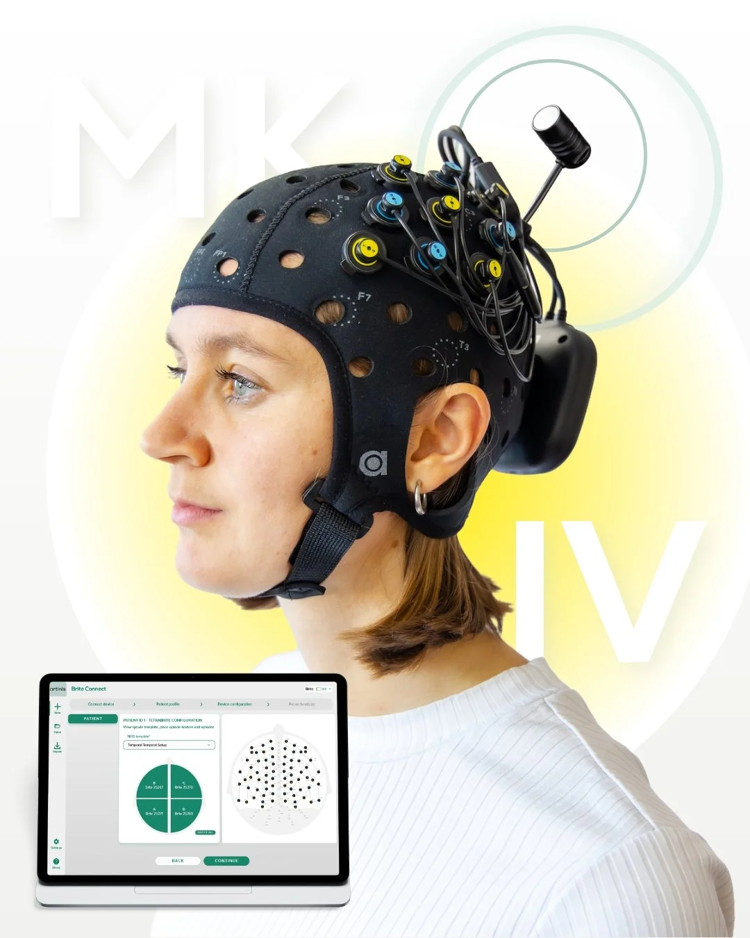
Measuring brain and muscle oxygenation at the same time with Artinis devices
As Artinis offers various devices that can be applied on any tissue, we provide different solutions to measure from brain and muscle at the same time.
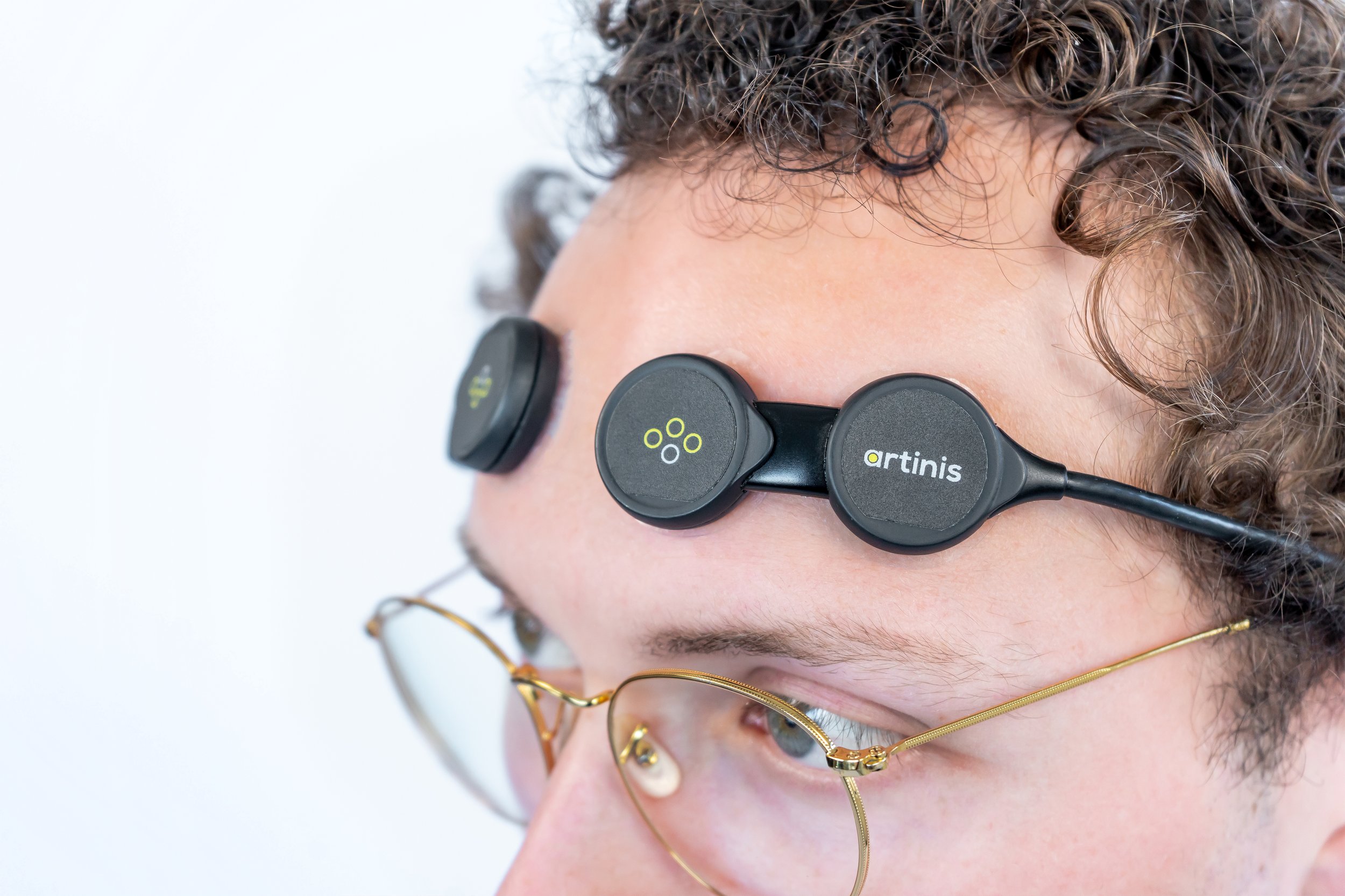
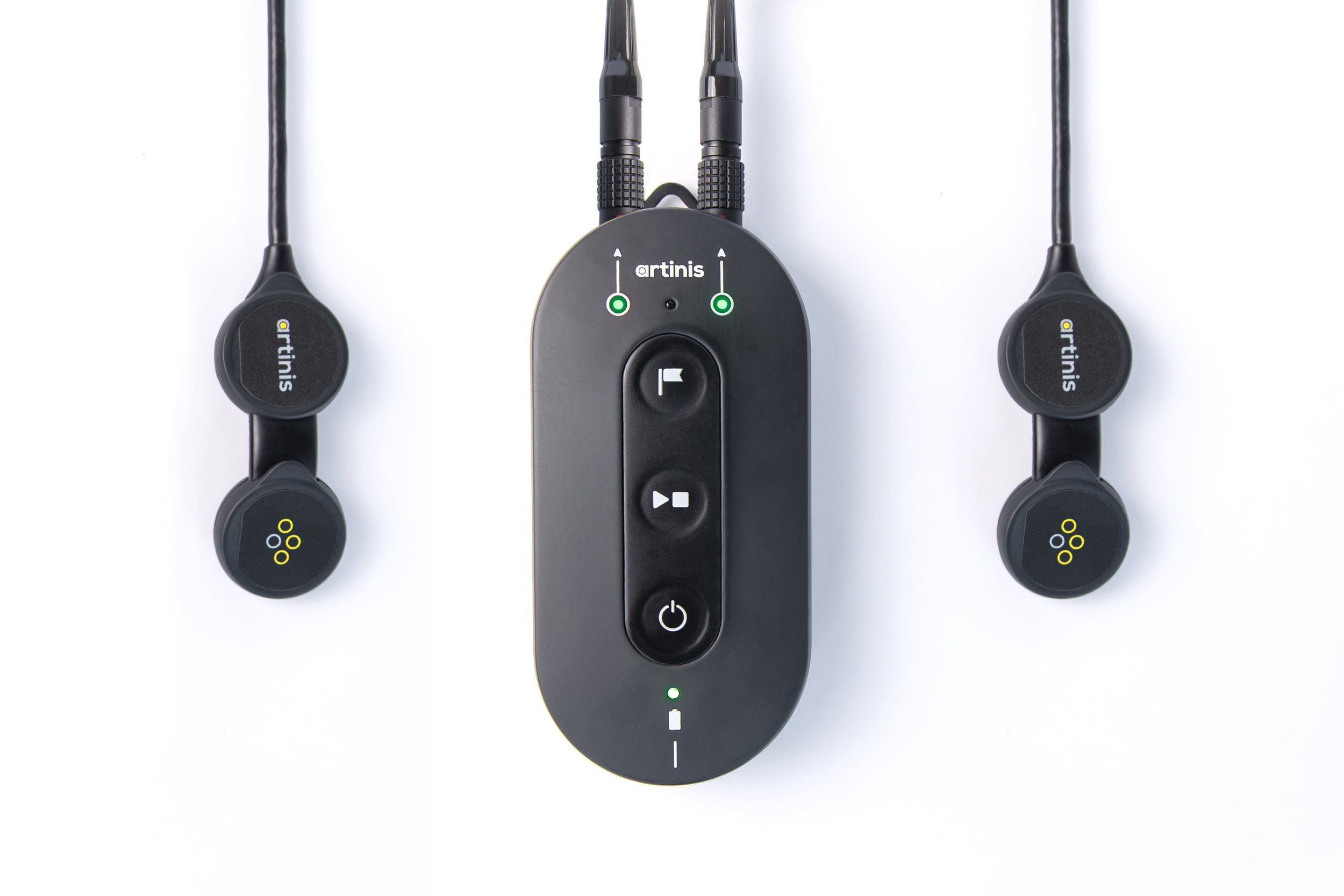
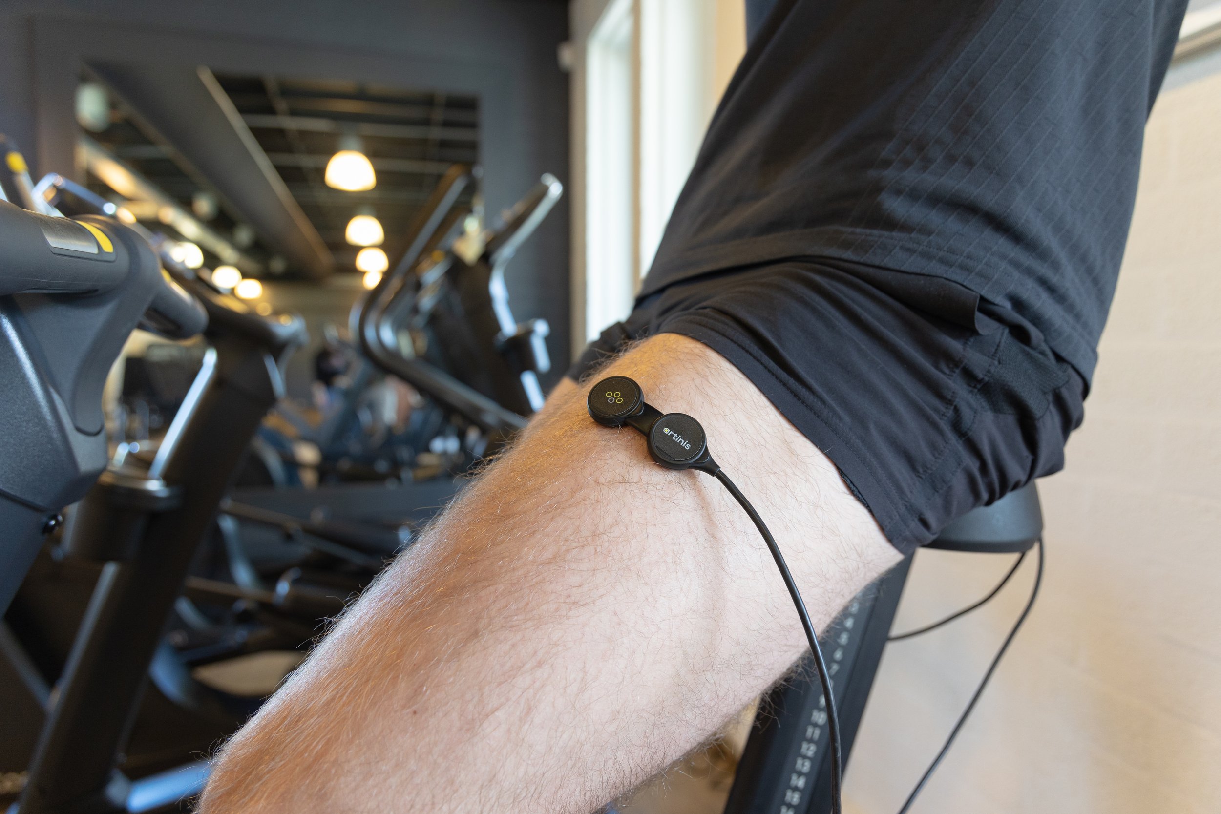
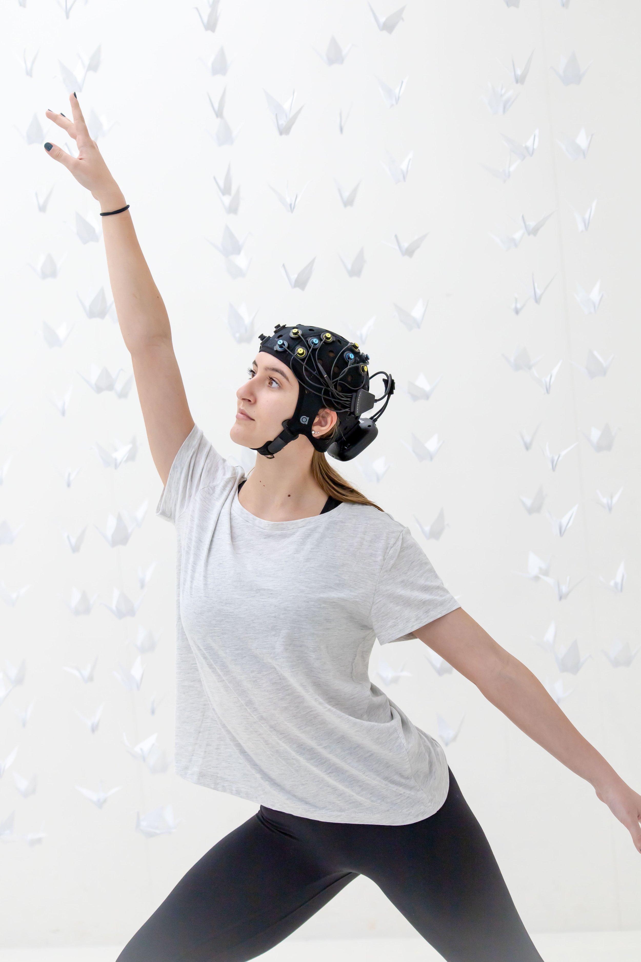
Our PortaLite MKII, which can connect two sensors to one control unit, offers the possibility to be applied on different sites at the same time. This enables measuring from brain and muscle simultaneously with only one device. The PortaLite is completely portable and wireless, leading to enhanced application possibilities in a variety of research fields. Further, it comes with superior features, such as a high sampling rate, short separation channels to measure from superficial tissue layers, or the possibility to add a 6-axis motion sensor, making it a reliable and robust tool to measure brain and muscle oxygenation in any kind of experiment.

When desiring enlarged brain coverage, or measuring from different muscles plus from the brain, other solutions are possible: Our software OxySoft allows to connect multiple devices at the same time, to ensure data synchronization while measuring from different locations. Therefore, devices to measure muscle oxygenation, such as the PortaMon and those to measure brain activity, for instance, the Brite Family devices, can be simultaneously used in one experiment, saving data in the same measurement file. Next to that, our devices are portable, wireless and lightweight, thus they can perfectly be applied in measurements that involve movement, or outside of typical lab settings.
Any questions left regarding simultaneous measurements of brain and muscle with our devices? Then feel free to reach out at askforinfo@artinis.com.


At Artinis, our mission is simple: make imaging easy, so researchers can focus on what matters, their research. Large-scale hyperscanning presented a clear challenge. We answered it the same way we always do: by removing the complexity and making it accessible.